Task 1. Examination of onion skin.
4. Draw a conclusion.
Answer. The skin of an onion consists of cells that fit tightly together.
Task 2. Examination of tomato cells (watermelon, apple).
1. Prepare a microslide of the fruit pulp. To do this, use a dissecting needle to separate a small piece of pulp from a cut tomato (watermelon, apple) and place it in a drop of water on a glass slide. Spread the dissecting needle in a drop of water and cover with a coverslip.
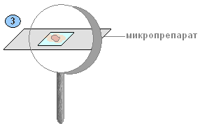
Answer. What to do. Take the pulp of the fruit. Place it in a drop of water on a glass slide (2).

2. Examine the microslide under a microscope. Find individual cells. Look at the cells at low magnification and then at high magnification.

Mark the color of the cell. Explain why the drop of water changed its color and why did this happen?
Answer. The color of the flesh cells of a watermelon is red, and that of an apple is yellow. A drop of water changes its color because it receives the cell sap contained in the vacuoles.
3. Draw a conclusion.
Answer. A living plant organism consists of cells. The contents of the cell are represented by semi-liquid transparent cytoplasm, which contains a denser nucleus with a nucleolus. The cell membrane is transparent, dense, elastic, does not allow the cytoplasm to spread, and gives it a certain shape. Some areas of the shell are thinner - these are pores, through which communication between cells occurs.
Thus, the cell is the structural unit of the plant
Even with the naked eye, or even better under a magnifying glass, you can see that the pulp of a ripe watermelon, tomato, or apple consists of very small grains or grains. These are cells - the smallest “building blocks” that make up the bodies of all living organisms.
What are we doing? Let's make a temporary microslide of a tomato fruit.
Wipe the slide and cover glass with a napkin. Use a pipette to place a drop of water on the glass slide (1).
What to do. Using a dissecting needle, take a small piece of fruit pulp and place it in a drop of water on a glass slide. Mash the pulp with a dissecting needle until you obtain a paste (2).

Cover with a cover glass and remove excess water with filter paper (3).
What to do. Examine the temporary microslide with a magnifying glass.
What we are seeing. It is clearly visible that the pulp of the tomato fruit has a granular structure (4).

These are the cells of the pulp of the tomato fruit.
What we do: Examine the microslide under a microscope. Find individual cells and examine them at low magnification (10x6), and then (5) at high magnification (10x30).

What we are seeing. The color of the tomato fruit cell has changed.
A drop of water also changed its color.
Conclusion: The main parts of a plant cell are the cell membrane, the cytoplasm with plastids, the nucleus, and vacuoles. The presence of plastids in the cell is a characteristic feature of all representatives of the plant kingdom.
Current page: 2 (book has 7 pages total) [available reading passage: 2 pages]
Biology is the science of life, of living organisms living on Earth.
Biology studies the structure and vital functions of living organisms, their diversity, and the laws of historical and individual development.
The area of distribution of life makes up a special shell of the Earth - the biosphere.
The branch of biology about the relationships of organisms with each other and with their environment is called ecology.
Biology is closely related to many aspects of human practical activity - agriculture, medicine, various industries, in particular food and light industries, etc.
Living organisms on our planet are very diverse. Scientists distinguish four kingdoms of living beings: Bacteria, Fungi, Plants and Animals.
Every living organism is made up of cells (with the exception of viruses). Living organisms eat, breathe, excrete waste products, grow, develop, reproduce, perceive environmental influences and react to them.
Each organism lives in a specific environment. Everything that surrounds a living being is called its habitat.
There are four main habitats on our planet, developed and inhabited by organisms. These are water, ground-air, soil and the environment inside living organisms.
Each environment has its own specific living conditions to which organisms adapt. This explains the great diversity of living organisms on our planet.
Environmental conditions have a certain impact (positive or negative) on the existence and geographical distribution of living beings. In this regard, environmental conditions are considered as environmental factors.
Conventionally, all environmental factors are divided into three main groups - abiotic, biotic and anthropogenic.
Chapter 1. Cellular structure of organisms
The world of living organisms is very diverse. To understand how they live, that is, how they grow, feed, and reproduce, it is necessary to study their structure.
In this chapter you will learn
About the structure of the cell and the vital processes occurring in it;
About the main types of tissues that make up organs;
About the structure of a magnifying glass, a microscope and the rules of working with them.
You will learn
Prepare microslides;
Use a magnifying glass and microscope;
In all organisms belonging to the same species, the number of chromosomes in cells is the same: in the house fly - 12, in Drosophila - 8, in corn - 20, in strawberries - 56, in crayfish - 116, in humans - 46, in chimpanzees , cockroach and pepper - 48. As you can see, the number of chromosomes does not depend on the level of organization.
Attention! This is an introductory fragment of the book.
If you liked the beginning of the book, then the full version can be purchased from our partner - the distributor of legal content, LitRes LLC.
3. Using the textbook, study the structure of hand-held and tripod magnifiers. Label their main parts in the pictures.
4. Examine pieces of fruit pulp under a magnifying glass. Sketch what you see. Sign the drawings.

5. After completing the laboratory work “The design of a microscope and methods of working with it” (see pp. 16-17 of the textbook), label the main parts of the microscope in the figure.

6. In the drawing, the artist mixed up the sequence of actions when preparing a microslide. Indicate with numbers the correct sequence of actions and describe the progress of preparing the microslide.
1) Place 1-2 drops of water on the glass.
2) Remove a small piece of transparent scale.
3) Place a piece of onion on the glass.
4) Cover with a cover slip and examine.
5) Stain the preparation with iodine solution.
6) Consider.
7. Using the text and pictures of the textbook (p. 2), study the structure of a plant cell, and then complete the laboratory work “Preparation and examination of a preparation of onion scale skin under a microscope.”

8. After completing the laboratory work “Plastids in the cells of the Elodea leaf” (see p. 20 of the textbook), sketch the structure of the cell of the Elodea leaf. Write captions for the drawing.

Conclusion: the cell has a complex structure: there is a nucleolus, cytoplasm, membrane, nucleus, vacuoles, pores, chloroplasts.
9. What color can plastids be? What other substances found in the cell give the plant organs different colors?
Green, yellow, orange, colorless.
10. Having studied paragraph 3 of the textbook, fill out the diagram “Cell life processes”.
Cell activity:
1) The movement of the cytoplasm - promotes the movement of nutrients in the cells.
2) Breathing – absorbs oxygen from the air.
3) Nutrition - from the intercellular spaces through the cell membrane they come in the form of nutrient solutions.
4) Reproduction - cells are capable of division, the number of cells increases.
5) Growth - cells increase in size.
11. Consider the division diagram of a plant cell. Use numbers to indicate the sequence of stages (stages) of cell division.

12. During life, changes occur in a cell.

Use numbers to indicate the sequence of changes from the youngest to the oldest cell.
3, 5, 1, 4, 2.
How does the youngest cell differ from the oldest cell?
The youngest cell has a nucleus, a nucleolus, and the oldest one does not.
13. What is the significance of chromosomes? Why is their number in a cell constant?
1) They transmit hereditary characteristics from cell to cell.
2) As a result of cell division, each chromosome copies itself. Two identical parts are formed.
14. Complete the definition.
A tissue is a group of cells that are similar in structure and perform the same functions.
15. Fill out the diagram.

16. Fill out the table.

17. Label the main parts of a plant cell in the picture.

18. What was the significance of the invention of the microscope?
The invention of the microscope was of great importance. With the help of a microscope, it became possible to see and examine the structure of the cell.
19. Prove that a cell is a living part of a plant.
A cell can: eat, breathe, grow, reproduce. And these are signs of living things.
Magnifier, microscope, telescope.
Question 2. What are they used for?
They are used to enlarge the object in question several times.
Laboratory work No. 1. Construction of a magnifying glass and using it to examine the cellular structure of plants.
1. Examine a hand-held magnifying glass. What parts does it have? What is their purpose?
A hand magnifying glass consists of a handle and a magnifying glass, convex on both sides and inserted into a frame. When working, the magnifying glass is taken by the handle and brought closer to the object at a distance at which the image of the object through the magnifying glass is most clear.
2. Examine with the naked eye the pulp of a semi-ripe tomato, watermelon, or apple. What is characteristic of their structure?
The pulp of the fruit is loose and consists of tiny grains. These are cells.
It is clearly visible that the pulp of the tomato fruit has a granular structure. The apple's pulp is slightly juicy, and the cells are small and tightly packed together. The pulp of a watermelon consists of many cells filled with juice, which are located either closer or further away.
Even with the naked eye, or even better under a magnifying glass, you can see that the flesh of a ripe watermelon consists of very small grains, or grains. These are cells - the smallest “building blocks” that make up the bodies of all living organisms. Also, the pulp of a tomato fruit under a magnifying glass consists of cells similar to rounded grains.
Laboratory work No. 2. The structure of a microscope and methods of working with it.
1. Examine the microscope. Find the tube, eyepiece, lens, tripod with stage, mirror, screws. Find out what each part means. Determine how many times the microscope magnifies the image of the object.

Tube is a tube that contains the eyepieces of a microscope. An eyepiece is an element of the optical system facing the eye of the observer, a part of the microscope designed to view the image formed by the mirror. The lens is designed to construct an enlarged image with accurate reproduction of the shape and color of the object of study. A tripod holds the tube with an eyepiece and objective at a certain distance from the stage on which the material being examined is placed. The mirror, which is located under the object stage, serves to supply a beam of light under the object in question, i.e., it improves the illumination of the object. Microscope screws are mechanisms for adjusting the most effective image on the eyepiece.
2. Familiarize yourself with the rules for using a microscope.
When working with a microscope, the following rules must be observed:
1. You should work with a microscope while sitting;
2. Inspect the microscope, wipe the lenses, eyepiece, mirror from dust with a soft cloth;
3. Place the microscope in front of you, slightly to the left, 2-3 cm from the edge of the table. Do not move it during operation;
4. Open the aperture completely;
5. Always start working with a microscope at low magnification;
6. Lower the lens to the working position, i.e. at a distance of 1 cm from the slide;
7. Set the illumination in the field of view of the microscope using a mirror. Looking into the eyepiece with one eye and using a mirror with a concave side, direct the light from the window into the lens, and then illuminate the field of view as much as possible and evenly;
8. Place the microspecimen on the stage so that the object being studied is under the lens. Looking from the side, lower the lens using the macroscrew until the distance between the lower lens of the lens and the microspecimen becomes 4-5 mm;
9. Look into the eyepiece with one eye and rotate the coarse aiming screw towards yourself, smoothly raising the lens to a position at which the image of the object can be clearly seen. You cannot look into the eyepiece and lower the lens. The front lens may crush the cover glass and cause scratches;
10. Moving the specimen with your hand, find the right place and place it in the center of the microscope’s field of view;
11. After finishing work with high magnification, set the magnification to low, raise the lens, remove the specimen from the work table, wipe all parts of the microscope with a clean napkin, cover it with a plastic bag and put it in a cabinet.
3. Practice the sequence of actions when working with a microscope.
1. Place the microscope with the tripod facing you at a distance of 5-10 cm from the edge of the table. Use a mirror to shine light into the opening of the stage.
2. Place the prepared preparation on the stage and secure the slide with clamps.
3. Using the screw, smoothly lower the tube so that the lower edge of the lens is at a distance of 1-2 mm from the specimen.
4. Look into the eyepiece with one eye without closing or squinting the other. While looking through the eyepiece, use the screws to slowly lift the tube until a clear image of the object appears.
5. After use, put the microscope in its case.
Question 1. What magnifying devices do you know?
Hand magnifier and tripod magnifier, microscope.
Question 2. What is a magnifying glass and what magnification does it provide?
A magnifying glass is the simplest magnifying device. A hand magnifying glass consists of a handle and a magnifying glass, convex on both sides and inserted into a frame. It magnifies objects 2-20 times.
A tripod magnifying glass magnifies objects 10-25 times. Two magnifying glasses are inserted into its frame, mounted on a stand - a tripod. A stage with a hole and a mirror is attached to the tripod.
Question 3. How does a microscope work?
Magnifying glasses (lenses) are inserted into the viewing tube, or tube, of this light microscope. At the upper end of the tube there is an eyepiece through which various objects are viewed. It consists of a frame and two magnifying glasses. At the lower end of the tube is placed a lens consisting of a frame and several magnifying glasses. The tube is attached to a tripod. An object table is also attached to the tripod, in the center of which there is a hole and a mirror under it. Using a light microscope, you can see an image of an object illuminated by this mirror.
Question 4. How to find out what magnification a microscope gives?
To find out how much the image is magnified when using a microscope, you need to multiply the number indicated on the eyepiece by the number indicated on the objective lens you are using. For example, if the eyepiece provides 10x magnification and the objective provides 20x magnification, then the total magnification is 10 x 20 = 200x.
Think
Why can't we study opaque objects using a light microscope?
The main principle of operation of a light microscope is that light rays pass through a transparent or translucent object (object of study) placed on the stage and hit the lens system of the objective and eyepiece. And light does not pass through opaque objects, and therefore we will not see an image.
Tasks
Learn the rules of working with a microscope (see above).
Using additional sources of information, find out what details of the structure of living organisms can be seen with the most modern microscopes.
The light microscope made it possible to examine the structure of cells and tissues of living organisms. And now, it has already been replaced by modern electron microscopes, which allow one to examine molecules and electrons. And an electron scanning microscope allows you to obtain images with a resolution measured in nanometers (10-9). It is possible to obtain data concerning the structure of the molecular and electronic composition of the surface layer of the surface under study.
Laboratory work No. 1
The device of magnifying devices
Target: study the structure of a magnifying glass and microscope and how to work with them.
Equipment: magnifying glass, microscope, tomato, watermelon, apple fruits .
Progress
The device of a magnifying glass and using it to examine the cellular structure of plants
1. Consider a hand-held magnifying glass. What parts does it have? What is their purpose?
2. Examine with the naked eye the pulp of a semi-ripe tomato, watermelon, or apple. What is characteristic of their structure?
3. Examine pieces of fruit pulp under a magnifying glass. Draw what you see in your notebook and sign the drawings. What shape do the fruit pulp cells have?
The device of a microscope and methods of working with it.
Examine the microscope. Find a tube, an eyepiece, screws, a lens, a tripod with a stage, a mirror. Find out what each part means. Determine how many times the microscope magnifies the image of the object.
Familiarize yourself with the rules for using a microscope.
Procedure for working with a microscope.
Place the microscope with the tripod facing you at a distance of 5–10 cm from the edge of the table. Use a mirror to direct light through the hole in the stage.
Place the prepared preparation on the stage and secure the slide with clamps.
Using the screws, smoothly lower the tube so that the lower edge of the lens is at a distance of 1 - 2 mm from the specimen.
After use, put the microscope in its case.
A microscope is a fragile and expensive device. You must work with it carefully, strictly following the rules.
Laboratory work No. 2
Target
Equipment
Progress
Stain the preparation with iodine solution. To do this, apply a drop of iodine solution to a glass slide. Use filter paper on the other side to pull off excess solution.
Laboratory work No. 3
Preparation of microslides and examination of plastids under a microscope in the cells of elodea leaves, tomato fruits, and rose hips.
Target: prepare a microslide and examine the plastids in the cells of an elodea, tomato and rose hip leaf under a microscope.
Equipment: microscope, elodea leaf, tomato and rose hips
Progress
Prepare a preparation of Elodea leaf cells. To do this, separate the leaf from the stem, place it in a drop of water on a glass slide and cover with a coverslip.
Examine the preparation under a microscope. Find chloroplasts in the cells.
Draw the structure of an Elodea leaf cell.
Prepare cell preparations of tomato, rowan, and rose hips. To do this, transfer a particle of pulp into a drop of water on a glass slide with a needle. Use the tip of a needle to separate the pulp into cells and cover with a coverslip. Compare the cells of the fruit pulp with the skin cells of the onion scales. Note the color of the plastids.
Sketch what you see. What are the similarities and differences between onion skin cells and fruit cells?
Laboratory work No. 2
Preparation and examination of a preparation of onion scale skin under a microscope
(structure of onion skin cells)
Target: study the structure of onion skin cells on a freshly prepared microslide.
Equipment: microscope, water, pipette, slide and cover glass, needle, iodine, bulb, gauze.
Progress
Look at Fig. 18 sequence of preparation of onion scale skin preparation.
Prepare the slide by wiping it thoroughly with gauze.
Use a pipette to place 1 – 2 drops of water onto a glass slide.
Using a dissecting needle, carefully remove a small piece of clear skin from the inside of the onion scale. Place a piece of peel in a drop of water and straighten it with the tip of a needle.
Cover the peel with a cover slip as shown in the picture.
Examine the prepared preparation at low magnification. Note which parts you see.
Stain the preparation with iodine solution. To do this, place a drop of iodine solution on a glass slide. Use filter paper on the other side to pull off excess solution.
Examine the colored preparation. What changes have occurred?
Examine the specimen at high magnification. Find a dark stripe surrounding the cell - the membrane, underneath it is a golden substance - the cytoplasm (it can occupy the entire cell or be located near the walls). The nucleus is clearly visible in the cytoplasm. Find the vacuole with cell sap (it differs from the cytoplasm in color).
Sketch 2 - 3 cells of onion skin. Label the membrane, cytoplasm, nucleus, vacuole with cell sap.
Laboratory work No. 4
Preparation of the preparation and examination under a microscope of the movement of the cytoplasm in the cells of the elodea leaf
Target: prepare a microscopic specimen of an elodea leaf and examine the movement of the cytoplasm in it under a microscope.
Equipment: freshly cut elodea leaf, microscope, dissecting needle, water, slide and cover glass.
Progress
Using the knowledge and skills acquired in previous lessons, prepare microslides.
Examine them under a microscope and note the movement of the cytoplasm.
Draw the cells, using arrows to show the direction of movement of the cytoplasm.
State your conclusion.
Laboratory work No. 5
Examination under a microscope of finished micropreparations of various plant tissues
Target: examine prepared micropreparations of various plant tissues under a microscope.
Equipment: micropreparations of various plant tissues, microscope.
Progress
Set up the microscope.
Under a microscope, examine ready-made micropreparations of various plant tissues.
Note the structural features of their cells.
Read P. 10.
Based on the results of studying the micropreparations and the text of the paragraph, fill out the table.
Laboratory work No. 6.
Structural features of mucor and yeast
Target: grow mucor mold and yeast, study their structure.
Equipment: bread, plate, microscope, warm water, pipette, slide, cover slip, wet sand.
Conditions for the experiment: heat, humidity.
Progress
Mucor mold
Grow white mold on bread. To do this, place a piece of bread on a layer of damp sand poured into a plate, cover it with another plate and place it in a warm place. After a few days, a fluff consisting of small threads of mucor will appear on the bread. Examine the mold with a magnifying glass at the beginning of its development and later, when black heads with spores form.
Prepare a microspecimen of the mold fungus mucor.
Examine the microscopic specimen at low and high magnification. Find mycelium, sporangia and spores.
Draw the structure of the mucor mushroom and label the names of its main parts.
Yeast structure
Dissolve a small piece of yeast in warm water. Pipette and place 1 – 2 drops of water with yeast cells on a glass slide.
Cover with a cover slip and examine the preparation using a microscope at low and high magnification. Compare what you see with Fig. 50. Find individual yeast cells, look at the outgrowths on their surface - buds.
Draw a yeast cell and label the names of its main parts.
Based on the research conducted, formulate conclusions.
Formulate a conclusion about the structural features of the mucor fungus and yeast.
Laboratory work No. 7
The structure of green algae
Target: study the structure of green algae
Equipment: microscope, slide, unicellular algae (Chlamydomonas, Chlorella), water.
Progress
Place a drop of “blooming” water on a microscope slide and cover with a coverslip.
Examine unicellular algae at low magnification. Look for Chlamydomonas (a pear-shaped body with a pointed front end) or Chlorella (a spherical body).
Pull off some of the water from under the cover glass with a strip of filter paper and examine the algae cell at high magnification.
Find the membrane, cytoplasm, nucleus, and chromatophore in the algae cell. Pay attention to the shape and color of the chromatophore.
Draw a cell and write the names of its parts. Check the correctness of the drawing using the drawings in the textbook.
State your conclusion.
Laboratory work No. 8.
The structure of moss, fern, horsetail.
Target: study the structure of moss, fern, horsetail.
Equipment: herbarium specimens of moss, fern, horsetail, microscope, magnifying glass.
Progress
STRUCTURE OF MOSS.
Consider a moss plant. Determine the features of its external structure, find the stem and leaves.
Determine the shape, location. Size and color of leaves. Examine the leaf under a microscope and sketch it.
Determine whether the plant has a branched or unbranched stem.
Examine the tops of the stem to find male and female plants.
Examine the spore box. What is the importance of spores in the life of mosses?
Compare the structure of moss with the structure of algae. What are the similarities and differences?
Write down your answers to the questions.
STRUCTURE OF THE SPORING TAIL
Using a magnifying glass, examine the summer and spring shoots of horsetail from the herbarium.
Find the spore-bearing spikelet. What is the significance of spores in the life of horsetail?
Sketch the horsetail shoots.
STRUCTURE OF A SPORING FERN
Study the external structure of the fern. Consider the shape and color of the rhizome: the shape, size and color of the fronds.
Examine the brown tubercles on the underside of the frond with a magnifying glass. What are they called? What develops in them? What is the importance of spores in the life of a fern?
Compare ferns with mosses. Look for similarities and differences.
Justify that fern belongs to higher spore plants.
What are the similarities between moss, fern, horsetail?
Laboratory work No. 9.
The structure of needles and conifer cones
Target: study the structure of conifer needles and cones.
Equipment: needles of spruce, fir, larch, cones of these gymnosperms.
Progress
Consider the shape of the needles and their location on the stem. Measure the length and pay attention to the color.
Using the description of the characteristics of coniferous trees presented below, determine which tree the branch you are considering belongs to.
The needles are long (up to 5 - 7 cm), sharp, convex on one side and rounded on the other, sitting in twos together...... Scots pine
The needles are short, hard, sharp, tetrahedral, sit singly, cover the entire branch...... ……………….Spruce
The needles are flat, soft, blunt, have two white stripes on the other side……………………………… Fir
The needles are light green, soft, sit in bunches, like tassels, fall off for the winter…………………………………….. Larch
Consider the shape, size, and color of the cones. Fill the table.
| Plant name | |||||||
| location | scale shape | density |
|||||
Separate one scale. Familiarize yourself with the location and external structure of the seeds. Why is the studied plant called gymnosperm?
Laboratory work No. 10.
Structure of flowering plants
Target: study the structure of flowering plants
Equipment: flowering plants (herbarium specimens), hand magnifying glass, pencils, dissecting needle.
| progress Consider a flowering plant. Find its root and shoot, determine their sizes and sketch their shape. Determine where the flowers and fruits are. Examine the flower, note its color and size. Examine the fruits and determine their quantity. Examine the flower. Find the pedicel, receptacle, perianths, pistils and stamens. Dissect the flower, count the number of sepals, petals and stamens. Consider the structure of the stamen. Find the anther and filament. Examine the anther and filament under a magnifying glass. It contains many pollen grains. Consider the structure of the pistil, find its parts. Cut the ovary crosswise and examine it under a magnifying glass. Find the ovule (ovule). What is formed from the ovule? Why are the stamens and pistil the main parts of a flower? Draw the parts of the flower and write their names? Questions to form a conclusion. What organs does a flowering plant consist of? What is a flower made of? |
The sizes of the cells are so small that it is impossible to examine them without special devices. Therefore, magnifying devices are used to study the structure of cells.
Magnifier- the simplest magnifying device. A magnifying glass consists of a magnifying glass, which is inserted into a frame with a handle for ease of use. Magnifiers come in handheld and tripod types.
A hand-held magnifying glass (Fig. 3, a) can magnify the object in question from 2 to 20 times.
Rice. 3. Hand-held (a) and tripod (b) magnifiers
A tripod magnifying glass (Fig. 3, b) magnifies the object 10-20 times. The rules for working with a magnifying glass are very simple: the magnifying glass must be brought to the object of study at a distance at which the image of this object becomes clear.
Using a magnifying glass, you can see the shape of fairly large cells, but it is impossible to study their structure.
(from the Greek micros - small and skopeo - I look) - an optical device for viewing in an enlarged form small objects that are not visible to the naked eye. With its help, they study, for example, the structure of cells.
A light microscope consists of a tube, or tube (from the Latin tube - tube). At the top of the tube there is an eyepiece (from the Latin oculus - eye). It consists of a frame and two magnifying glasses. At the lower end of the tube there is a lens (from the Latin objectum - object), consisting of a frame and several magnifying glasses. The tube is attached to a tripod. The tube is raised and lowered using screws. There is also a stage on the tripod, in the center of which there is a hole and a mirror underneath it. The object examined on the slide is placed on the stage and secured to it using clamps (Fig. 4).

Rice. 4. Light microscope
The main principle of operation of a light microscope is that light rays pass through a transparent (or translucent) object of study, which is located on the stage, and fall on a system of objective lenses and an eyepiece, which magnify the image. Modern light microscopes can magnify images up to 3,600 times.
To find out how much the image is magnified when using a microscope, you need to multiply the number indicated on the eyepiece by the number indicated on the objective lens you are using. For example, if the number 8 is on the eyepiece and 20 on the lens, then the magnification factor will be 8 x 20 = 160.
Answer the questions
- What instruments are used to study cells?
- What are magnifying glasses and how much magnification can they provide?
- What parts does a light microscope consist of?
- How to determine the magnification given by a light microscope?
New concepts
Cell. Magnifier. Light microscope: eyepiece, lens.
Think!
Why can't we study opaque objects using a light microscope?
My laboratory
Some cells can be seen with the naked eye. These are the cells of the pulp of the fruits of watermelon, tomato, nettle fiber (their length reaches 8 cm), the yolk of a chicken egg - one large cell.

Rice. 5. Tomato cells under a magnifying glass
Examining the cellular structure of plants using the moon
- Examine with the naked eye the pulp of tomato, watermelon, and apple fruits. What is characteristic of their structure?
- Examine pieces of fruit pulp under a magnifying glass. Compare what you see with Figure 5, sketch it in your notebook, and sign the drawings. What shape do the fruit pulp cells have?
The structure of a light microscope and methods of working with it
- Study the structure of the microscope using Figure 4. Find the tube, eyepiece, lens, tripod with stage, mirror, screws. Find out what each part means.
- Familiarize yourself with the rules of using a microscope.
- Practice the procedure for working with a microscope!
Rules for working with a microscope
- Place the microscope with the tripod facing you at a distance of 5-10 cm from the edge of the table. Use a mirror to shine light into the opening of the stage.
- Place the slide with the prepared preparation on the stage. Secure the slide with clamps.
- Using the screw, smoothly lower the tube so that the lower edge of the lens is at a distance of 1-2 mm from the specimen.
- Look into the eyepiece with one eye without closing or squinting the other. While looking through the eyepiece, use the screws to slowly lift the tube until a clear image of the object appears.
- After use, put the microscope in its case.
- A microscope is a fragile and expensive device: you must work with it carefully, strictly following the rules.
The first microscopes with two lenses were invented at the end of the 16th century. However, it was not until 1665 that the Englishman Robert Hooke used the microscope he had improved to study organisms. Examining a thin section of cork (the bark of a cork oak tree) through a microscope, he counted up to 125 million pores, or cells, in one square inch (2.5 cm). Hooke discovered the same cells in the core of elderberry and the stems of various plants. He gave them the name “cells” (Fig. 6).

Rice. 6. Microscope of R. Hooke and view of cork cells according to his own drawing
At the end of the 17th century. Dutchman Antonie van Leeuwenhoek designed a more advanced microscope, providing magnification up to 270 times (Fig. 7). With his help, he discovered microorganisms. Thus began the study of the cellular structure of organisms.
Rice. 7. Microscope by A. Leeuwenhoek.
A magnifying glass (a) is attached to the upper part of the metal plate. The observed object was located at the tip of a sharp needle (b). The screws served for focusing.
While studying plant science, botany and carpology in practice, it is interesting to touch upon the topic of the apple tree and its multi-seeded, indehiscent fruits, which humans have eaten since ancient times. There are many varieties, the most common type is “domestic”. It is from it that manufacturers all over the world make canned food and drinks. Looking at the apple under microscope one can note the similarity of the structure with a berry, which has a thin shell and a juicy core and contains multicellular structures - seeds.
 The apple is the final stage of flower development on the apple tree, occurring after double fertilization. Formed from the ovary of the pistil. From it the pericarp (or pericarp) is formed, which performs a protective function and serves for further reproduction. It, in turn, is divided into three layers: exocarp (outer), mesocarp (middle), endocarp (inner).
 Analyzing the morphology of apple tissue at the cell level, we can distinguish the main organelles:
- Cytoplasm is a semi-liquid medium of organic and inorganic substances. For example, salts, monosaccharides, carboxylic acids. It combines all components into a single biological mechanism, providing endoplasmic cyclosis.
- A vacuole is an empty space filled with cell sap. It organizes salt metabolism and serves to remove metabolic products.
- The nucleus is the carrier of genetic material. It is surrounded by a membrane.
Methods of observation apple under a microscope:

- Transmitted lighting. The light source is located under the test drug. The microsample itself must be very thin, almost transparent. For these purposes, a slice is prepared using the technology described below.
Preparation of a microslide of apple pulp:
- Use a scalpel to make a rectangular incision and carefully remove the skin with tweezers;
- Using a medical dissecting needle with a straight tip, transfer a piece of flesh to the center of the slide;
- Using a pipette, add one drop of water and a dye, for example, a solution of brilliant green;
- Cover with a coverslip;
It is best to start microscopying with a low magnification of 40x, gradually increasing the magnification to 400x (maximum 640x). The results can be recorded digitally by displaying the image on a computer screen using an eyepiece camera. It is usually purchased as an additional accessory and is characterized by the number of megapixels. It was used to take the photos presented in this article. To take a photo, you need to focus and press the virtual photo button in the program interface. Short videos are made in the same way. The software includes functionality that allows linear and angular measurements of areas of particular interest to the observer.
If you examine the pulp of a tomato or watermelon with a microscope magnifying approximately 56 times, round transparent cells are visible. In apples they are colorless, in watermelons and tomatoes they are pale pink.
1050;the cells in the “mush” lie loosely, separated from each other, and therefore it is clearly visible that each cell has its own shell, or wall.
Conclusion: A living plant cell has:
1. Living contents of the cell. (cytoplasm, vacuole, nucleus)
2. Various inclusions in the living contents of the cell.
#1086;deposition of reserve nutrients: protein grains, drops of oil, starch grains.)
3. Cell membrane, or wall. (It is transparent, dense, elastic, does not allow the cytoplasm to spread, and gives the cell a certain shape.)
Magnifier, microscope, telescope.
Question 2. What are they used for?
They are used to enlarge the object in question several times.
Laboratory work No. 1. Construction of a magnifying glass and using it to examine the cellular structure of plants.
1. Examine a hand-held magnifying glass. What parts does it have? What is their purpose?
A hand magnifying glass consists of a handle and a magnifying glass, convex on both sides and inserted into a frame. When working, the magnifying glass is taken by the handle and brought closer to the object at a distance at which the image of the object through the magnifying glass is most clear.
2. Examine with the naked eye the pulp of a semi-ripe tomato, watermelon, or apple. What is characteristic of their structure?
The pulp of the fruit is loose and consists of tiny grains. These are cells.
It is clearly visible that the pulp of the tomato fruit has a granular structure. The apple's pulp is slightly juicy, and the cells are small and tightly packed together. The pulp of a watermelon consists of many cells filled with juice, which are located either closer or further away.
3. Examine pieces of fruit pulp under a magnifying glass. Draw what you see in your notebook and sign the drawings. What shape do the fruit pulp cells have?
Even with the naked eye, or even better under a magnifying glass, you can see that the flesh of a ripe watermelon consists of very small grains, or grains. These are cells - the smallest “building blocks” that make up the bodies of all living organisms. Also, the pulp of a tomato fruit under a magnifying glass consists of cells similar to rounded grains.
Laboratory work No. 2. The structure of a microscope and methods of working with it.
1. Examine the microscope. Find the tube, eyepiece, lens, tripod with stage, mirror, screws. Find out what each part means. Determine how many times the microscope magnifies the image of the object.

Tube is a tube that contains the eyepieces of a microscope. An eyepiece is an element of the optical system facing the eye of the observer, a part of the microscope designed to view the image formed by the mirror. The lens is designed to construct an enlarged image with accurate reproduction of the shape and color of the object of study. A tripod holds the tube with an eyepiece and objective at a certain distance from the stage on which the material being examined is placed. The mirror, which is located under the object stage, serves to supply a beam of light under the object in question, i.e., it improves the illumination of the object. Microscope screws are mechanisms for adjusting the most effective image on the eyepiece.
2. Familiarize yourself with the rules for using a microscope.
When working with a microscope, the following rules must be observed:
1. You should work with a microscope while sitting;
2. Inspect the microscope, wipe the lenses, eyepiece, mirror from dust with a soft cloth;
3. Place the microscope in front of you, slightly to the left, 2-3 cm from the edge of the table. Do not move it during operation;
4. Open the aperture completely;
5. Always start working with a microscope at low magnification;
6. Lower the lens to the working position, i.e. at a distance of 1 cm from the slide;
7. Set the illumination in the field of view of the microscope using a mirror. Looking into the eyepiece with one eye and using a mirror with a concave side, direct the light from the window into the lens, and then illuminate the field of view as much as possible and evenly;
8. Place the microspecimen on the stage so that the object being studied is under the lens. Looking from the side, lower the lens using the macroscrew until the distance between the lower lens of the lens and the microspecimen becomes 4-5 mm;
9. Look into the eyepiece with one eye and rotate the coarse aiming screw towards yourself, smoothly raising the lens to a position at which the image of the object can be clearly seen. You cannot look into the eyepiece and lower the lens. The front lens may crush the cover glass and cause scratches;
10. Moving the specimen by hand, find the desired location and place it in the center of the microscope’s field of view;
11. After finishing work with high magnification, set the magnification to low, raise the lens, remove the specimen from the work table, wipe all parts of the microscope with a clean napkin, cover it with a plastic bag and put it in a cabinet.
3. Practice the sequence of actions when working with a microscope.
1. Place the microscope with the tripod facing you at a distance of 5-10 cm from the edge of the table. Use a mirror to shine light into the opening of the stage.
2. Place the prepared preparation on the stage and secure the slide with clamps.
3. Using the screw, smoothly lower the tube so that the lower edge of the lens is at a distance of 1-2 mm from the specimen.
4. Look into the eyepiece with one eye without closing or squinting the other. While looking through the eyepiece, use the screws to slowly lift the tube until a clear image of the object appears.
5. After use, put the microscope in its case.
Question 1. What magnifying devices do you know?
Hand magnifier and tripod magnifier, microscope.
Question 2. What is a magnifying glass and what magnification does it provide?
A magnifying glass is the simplest magnifying device. A hand magnifying glass consists of a handle and a magnifying glass, convex on both sides and inserted into a frame. It magnifies objects 2-20 times.
A tripod magnifying glass magnifies objects 10-25 times. Two magnifying glasses are inserted into its frame, mounted on a stand - a tripod. A stage with a hole and a mirror is attached to the tripod.
Question 3. How does a microscope work?
Magnifying glasses (lenses) are inserted into the viewing tube, or tube, of this light microscope. At the upper end of the tube there is an eyepiece through which various objects are viewed. It consists of a frame and two magnifying glasses. At the lower end of the tube is placed a lens consisting of a frame and several magnifying glasses. The tube is attached to a tripod. An object table is also attached to the tripod, in the center of which there is a hole and a mirror under it. Using a light microscope, you can see an image of an object illuminated by this mirror.
Question 4. How to find out what magnification a microscope gives?
To find out how much the image is magnified when using a microscope, you need to multiply the number indicated on the eyepiece by the number indicated on the objective lens you are using. For example, if the eyepiece provides 10x magnification and the objective provides 20x magnification, then the total magnification is 10 x 20 = 200x.
Think
Why can't we study opaque objects using a light microscope?
The main principle of operation of a light microscope is that light rays pass through a transparent or translucent object (object of study) placed on the stage and hit the lens system of the objective and eyepiece. And light does not pass through opaque objects, and therefore we will not see an image.
Tasks
Learn the rules of working with a microscope (see above).
Using additional sources of information, find out what details of the structure of living organisms can be seen with the most modern microscopes.
The light microscope made it possible to examine the structure of cells and tissues of living organisms. And now, it has been replaced by modern electron microscopes, which allow us to examine molecules and electrons. And an electron scanning microscope allows you to obtain images with a resolution measured in nanometers (10-9). It is possible to obtain data concerning the structure of the molecular and electronic composition of the surface layer of the surface under study.